During this case study we demonstrate the development of in-cell competition assays to identify compounds that interact with target kinases. Measuring potency and residency time on the binding site within a live cell environment.
Challenges
What parameters are important during the design-make-test-understand cycle of compound progression when examining interaction to a kinase target? Binding affinity and selectivity are used for Structure Activity Relationships (SAR) progression but what about the kinetics of compound binding?
The association (kon) and dissociation (koff) rates of compounds are important considerations. Is 1 nM binding and 60-minute occupancy ‘better’ than 100 nM binding and 8-hour occupancy in living cells? How would occupancy time influence off-target effects, and could these be removed by studying binding kinetics?
When choosing which compounds to progress this may influence your SAR decisions. For example, if you are developing a PROTAC (PRoteOlysis Targeted Chimera) for targeted protein degradation (TPD) is it better to have a target warhead with a high residency time to allow maximum occupancy time for target ubiquitination? Does this approach affect the characteristic ‘hook effect’ observed with PROTAC’s and would this be more efficacious?
Using Promega Target Engagement reagents we have developed assays designed to study the interaction (kon and koff) of compounds on kinase targets importantly within living cells at physiological ATP concentrations, studying the rate of association and dissociation of compounds for kinase targets in living cells.
What We Did...
How the Technology Works Nanoluc® & NanoBRET™
The technology uses Bioluminescence Energy Transfer (BRET) to measure compound engagement with the kinase target. The full-length target kinase cDNA connected to the Nanoluc enzyme cDNA on a single plasmid are transiently transfected into cells for 24-hours. When expressed, the kinase protein is connected to the luminescence enzyme (NanoLuc).
In the presence of a cell permeable Nanoluc substrate, photons are generated and emitted at 460 nm. When cells are treated with a cell-permeable fluorescently tagged tracer, binding of the tracer to the kinase brings the tag into close proximity with NanoLuc and the photons excite the fluorescent tag resulting in fluorescence emission at 600+ nm and generating BRET.
Compound engagement is measured in a competitive format of tracer versus compound. Binding of the test compound results in a loss of NanoBRET signal between the target protein and the tracer inside living intact cells.
For analysis of target engagement by a test compound, cells are treated with a fixed concentration of NanoBRET tracer that is near the EC50 value of the NanoBRET tracer dose response curve. To determine test compound affinity, cells are titrated with varying concentrations of the test compound in the presence of a fixed concentration (EC50–EC80) of tracer.

Figure 1. How the technology works. The target plus Nanoluc protein is over-expressed in cells, the fluorescence tagged small molecule tracer binds to the target resulting in BRET in the presence of Nanoluc substrate. Competing compounds displace the tracer and the NanoBRET signal decreases. Data is expressed as a ratio of excitation photons at 460 nm divided by fluorescence emission at 600+ nm.
Kinase Tracer Affinity Determination
We evaluated the technology using four kinase targets; ABL-1, FGR, EPHA8 and DDR1. For each kinase, the appropriate tracer (K4 in this case) was tested as a dose-response to determine the EC50 for each target and was competed off the target with molar excess of unlabelled tracer. By using fixed concentrations of tracer, we were then able to perform dose-response curves using the unlabelled tracer, to determine the affinity of the unlabelled tracer compound (see table 1 below).
Tracer Concentration (µM)
Kinase
3
1
0.33
0.11
0.037
0.012
0.0041
ABL
–
22
10
8
8
4
–
FGR
49
33
20
31
–
–
–
EPHA8
–
–
14
6
3
2
2
DDR-1
–
–
8
5
4
3
2


Figure 3. Binding activity of each compound was determined in a living cell competition assay. Cells were transfected for 24-hours with each of four kinase; ABL, FGR, EPHA8 and DDR-1. Cells were treated with exemplar kinase compounds including dasatinib, nilotinib, foretinib and ponatinib as a dose-response for each compound competed against a fixed concentration of fluorescent tracer K4 at the concentration depicted in table 2 below. As these compounds are pan-kinase binders, all compete off the tracer in a dose-dependent manner except nilotenib and fortenib from the FGR kinase.
ABL
EPHA8
FGR
DDR-1
Dasatinib
11
<1
45
4
Nilotinib
340
164
IA
3
Foretinib
~2000
107
IA
12
Ponatinib
480
27
~600
67
Validation Prior to High Throughput Screening (HTS) on One Kinase and Selectivity Screening Against Others
A subset of 80 random compounds were arrayed onto a 96 well plate and discrete wells were spiked with an active compound. All compounds were tested at 10 µM on each of the four kinase in the target engagement assay.
As expected, most compounds showed a distribution centered around zero percent inhibition (no binding) and all spiked active compounds were detected in each plate tested. The Z factor for each plate was above 0.7. Data suggests that all four kinase assays could be used as a high throughput screen to detect compounds that binding to the kinase target in this living cell competition assay.

Figure 4. Distribution of activity of 80 random compounds screened on each of the four kinase targets (ABL, FGR, EPHA8 and DDR-1) within the target engagement assay. The spike around 60 to 70% represents the activity of the spiked control compound in each case at 10 µM.
HTS Screening Approach
Large Scale Transfection Followed by Cryo-preservation and Subsequent use of Vials for Screening Purposes
To dissociate culture plus transfection from the screening stage, in an attempt to increase the day-to-day consistency and throughput of the assay for HTS purposes, HEK-293 cells were transiently transfected with a construct containing DDR-1 Nanoluc and cultured for 24-hours.
Cells were then divided into two aliquots, one was cryo-preserved, while the other was evaluated for binding using dasatinib and tracer K4. Frozen cells were defrosted and also assayed using dasatinib and tracer K4. The IC50 for dasatinib between fresh and frozen cells was very similar (see below).
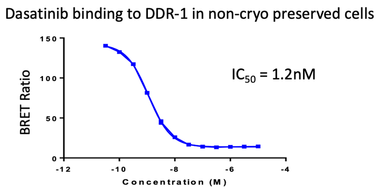
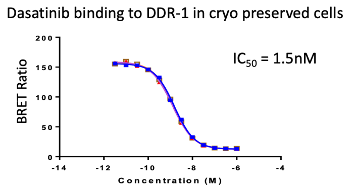
Figure 5. Evalaution of cryo-preservation on the ability to use the cells straight from the cryostore. Cells transfected with cDNA for DDR-1 were cryopreserved then defrosted and used in the same day (following a 2-hour recovery period) to examine the competitive binding of dasatinib.
Both frozen and fresh cells showed similar IC50’s, demonstrating that this format could be used for HTS direct to assay purposes, improving throughput and the efficiency of selectivity testing of a single compound against many kinase targets as we can have aliquots of frozen transfected cells available for kinase selectivity testing.
Kinetics of Binding: On- & Off-rates of Compounds Inside Cells
By studying both the association and dissociation rate of compounds from each of the target kinase, it is possible to consider introducing kinetic binding parameter evaluation during compound SAR development. This can be measured intracellular at physiological ATP and pH when the cells are in culture within the assay. Below we have examined the association rate of the compounds after the cells have been ‘soaked’ in tracer. The time dependent decline in BRET signal is proportional to the rate of association of the compound.

In the reverse experiment, cells were ‘soaked’ for 2-hours in compound, then tracer added at a fixed concentration. In this case the tracer displaces the compound from the binding site and an increase in BRET is observed. The orange line represents the binding of the tracer to the kinase in the absence of compound (DMSO alone). In this format it is possible to observe covalent compounds (see FGR) that cannot be competed by the tracer from the binding pocket.

Summary
We have developed high throughput screening assays in living cells looking at the competition of compounds to displace the fluorescent tracer and displaying a good assay quality and reproducibility (Z factor = 0.7 and above).
By using cryo-preserved cells, we can simply and efficiently perform high throughput screening using the same transfected batch of cells plus selectivity screening in which we have a number of vials of cells expressing separate kinase and, taking a single compound, can test for binding against many kinase targets.
In addition, we can determine the binding (as IC50’s) of exemplar compounds such as dasatinib, nilotibin, foretinib and ponatinib to ABL, FGR, EPHA8 and DDR-1 kinases. Furthermore, we can measure both the association constant (kon) and dissociation (koff) for compounds within living cells at physiological ATP concentration and pH.
These assays can be used to examine binding and kinetics for individual kinase targets and off-target kinase toxicity testing. Finally, the cDNA construct can be designed to work within a CRISPR Cas9 system to replace the endogenous kinase in different cell backgrounds. This technology represents a significant step forward for SAR screening as compound kinetics of binding can, in addition to potency, be used to rank compounds in projects within a physiological environment expressing the full-length kinase target.