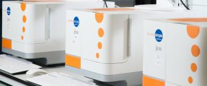
We demonstrate the use of the JESS automated western blot platform, coupled with “Felix,” a robotic liquid handler from Analytic Jena. This combination automates the process, ensuring a truly reproducible and robust Western blotting screening assay.
Separating and quantifying specific proteins within cells is crucial for studying normal function and disease. Drug discovery approaches, like PROTAC or biomarker detection, depend on accurate protein quantitation. Traditional western blotting is limited by throughput, detection sensitivity, and reproducibility. These limitations are critical when screening novel compounds.
Challenges
The Simple-Western™ JESS technology from Biotechne is a capillary-based automated western blot instrument. It can screen 24 cell lysate samples, delivering results in about five hours. This is significantly faster than traditional western blotting, which takes 24-48 hours from lysate preparation to data visualization. Automation of protein separation and immunodetection eliminates error-prone steps found in traditional methods. The associated software rapidly processes quantifiable data.
Our aim was to further simplify the Simple-Western™ procedure, enhancing reproducibility, flexibility, and the ability to multiplex. This maximizes the design-make-test cycle turnaround for scientists. We tested the technology using THAL-SNS-032, a well-studied PROTAC, to examine its capability to degrade CDK9.
What We Did...
When constructing a new JESS assay, antibodies undergo optimization for each target protein and cell line. This ensures antibody saturation and concentration-dependent signal linearity.
In this study, we optimized the anti-CDK9 antibody and the loading control antibody (β-Actin) using the CyBio FeLiX automated dispenser. Employing a stock cell lysate (HEK293T), the CyBio FeLiX conducted a serial dilution of the lysate and prepared the proprietary JESS plate with varying antibody dilutions.
This automation eliminated the hands-on component typically required, enhancing the accuracy of dispensing, sample tracking, and speed (see Figure 1A). Data analysis involved fitting peaks (see Figure 1A) and generating a pseudo-image. Our findings demonstrated protein concentration-dependent detection of CDK9 and β-Actin (see Figure 1B).

Figure 1A. Schematic of CyBio FeLiX antibody optimization JESS plate preparation.
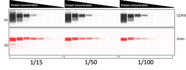
Figure 1B. Pseudo-image of CDK9 and β-Actin optimization with 8 protein concentrations and 3 antibody dilution factors, all multiplexed within the same lanes. CDK9 was detected using a horse radish peroxidase labelled secondary antibody, actin was detected using a near-infra red labelled secondary antibody.
It is important that peak signal demonstrates a linear correlation with protein concentration (Figure. 2) and that the signal remains consistent across antibody dilutions to ensure saturation (Figure. 2.).

Figure. 2. Graph (left) demonstrating linear relationship between protein concentration and CDK9 signal intensity. Graph (right) showing antibody saturation with a constant signal intensity across varying protein concentrations.
Cells were seeded and treated with serially diluted SNS-THAL-032 before being collected, washed with PBS, then lysed with RIPA lysis buffer (Figure. 3).

Figure 3. Schematic of how JESS samples were generated
To ascertain the protein concentration of assay samples, the FeLiX user loaded the lysates onto the deck, and the CyBio FeLiX automatically diluted the samples and added assay reagents to perform a BCA protein assay (refer to Figure 4).
The BCA assay serves to ensure that sample protein concentrations fall within the linear range of the antibody. This precaution helps prevent instances of camera saturation (on JESS) or substrate depletion (such as horse radish peroxidase). By eliminating any ‘hands-on’ requirements, this process enhances reproducibility.

Figure 4. Schematic of automated BCA Assay to measure sample protein concentration.
A protein standard curve is generated, enabling the extrapolation of unknown sample concentrations to be calculated using BCA reagents and absorbance at 562 nm as the readout. Therefore, ensuring reproducibility of the standard curve and accuracy between replicates is of vital importance (see Figure 5).

Figure. 5. A BSA standard curve was generated on three separate occasions by the CyBio FeLiX, demonstrating the reproducibility of the dispensing system.
Once all the optimization steps have been completed, CDK9 protein levels within the treated cells can be examined. The CyBio FeLiX performed dilution of the protein lysates to their optimal concentration before automatically loading them and all additional JESS reagents onto the JESS plate. To assess the reproducibility of JESS plate preparation, three biological replicates were conducted (refer to Figure 6).

Figure 6. Schematic of CyBio FeLiX JESS plate preparation. This format of plate and identical samples were used to generate three technical replicates (three separate experiments on the JESS with the same samples in three separate runs).
SNS-THAL-032 demonstrated potent and reproducible dose dependent degradation of CDK9 with an average EC50 of 16.1 nM ± 7.6 nM (Figure. 7.A and 7.B).


Figure. 7.A. Representative JESS pseudo-images of CDK9 dose dependent degradation by SNS-THAL-032. CDK9 was detected in the luminescence channel using a HRP tagged secondary antibody while Actin was detected using a near-infra red tagged secondary antibody.
Figure. 7.B. EC50 curves of three biological replicates showing degradation of CDK9 by THAL-SNS-032 (error bars represent SD) of three technical replicates.
When comparing CyBio FeLiX to manually prepared historical JESS data (see Figure 8), the results showcased the capability to replicate the data using a programmable liquid dispenser. This not only saves up to 60 minutes of bench time per JESS run but also enables scientists to multiplex their time. Additionally, it ensures consistency in automated dispensing.
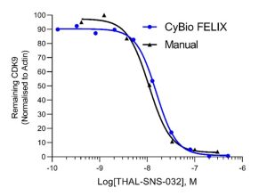
Figure. 8. EC50 curves for the averaged data from three CyBio FeLiX prepared JESS plates and a single manually prepared JESS plate.
Summary
The collaboration between the Simple-Western™ JESS expertise at Charnwood Discovery and the liquid handling automation expertise at Analytik-Jena has resulted in the development of a fully automated western blotting method for JESS plate preparation.
Utilizing the CyBio FeLiX, samples were processed from the initial BCA protein assay through to quantifiable results from the JESS instrument. This streamlined process reduces bench-time by at least 60 minutes per scientist per JESS run. For advanced users of the Simple-Western™ JESS technology, this translates to a potential saving of up to 45 hours of bench-time per week.
Furthermore, implementation of automation has led to a decrease in the variability observed in pipetting between scientists, thereby enhancing confidence in sample management and location.