The altered pH environment found within the center of a spheroid often can change the way a drug interacts with the target. This project outlines how our bioscience department developed an assay to test the cell viability in the presence of the compound.
Challenges
Examine the activity of test molecules in spheroids generated from cancer cell lines.
A differential pH was tested for maximum cyto-toxic effect, not observed in a 2D monolayers, but localized in the center of the spheroid, and different to that on the periphery, allowing more significant toxicity of the test molecules through a proprietary mechanism.
What We Did...
Using a 3D spheroid approach, we monitored compound potency and efficacy in either an imaging-based assay or using commercially available assays such as the CellTiter Glo® 3D viability assay (Promega). Here, we test two spheroid cultures derived from either HCT116 (human colorectal carcinoma) or U87 (human glioblastoma) cell lines. We have used several readouts to generate a pharmacological profile in each spheroid type. In addition to the test molecules, nocodazole, pacilitaxel, mitomycin C, WYE 687, staurosporine and camptothecin were used as positive control compounds.
Cell Viability
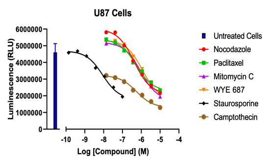
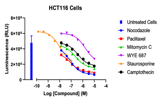
Figure 1. Spheroids were formed in ultra-lower attachment plates for 4-days to optimize cell compaction prior to compound treatment. To assess viability, spheroids constructed from U87 (left) and HCT116 (right) cells were cultured and treated with a dose-response to compound. Each cell line showed a dose-dependent decrease in viability, as determined by ATP concentration.
Nocodazole
Paclitaxel
Mitomycin C
WYE 687
Sturoprine
Camptothecin
U87
416 nM
535 nM
935 nM
1165 nM
9 nM
553 nM
HCT116
78 nM
84 nM
229 nM
1622 nM
10 nM
376 nM
Table 1. Estimated IC50 values determined in the CellTiter Glo® 3D Viability assay in spheroids derived from U87 or HCT116 cells.
Growth Inhibition
Initially, spheroid sizes showed a decrease due to cell compaction. Thereafter, in the absence of compound, spheroids grew in a linear manner up to day 8. Compounds induced a dose-dependent inhibition of growth, as characterized by a reduction in the spheroid size, consistently seen in the U87 cell line. In comparison, paclitaxel and nocodazole induced a looser cytoskeletal organization in HCT116 spheroids at higher concentrations.
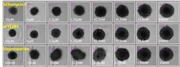
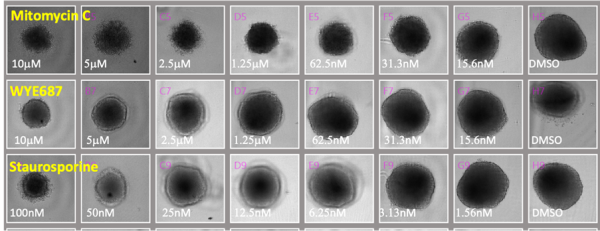
Figure 2: Brightfield images of spheroids U87 cells (top panel) or HCT116 cells (bottom panel).
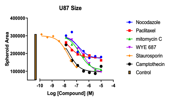
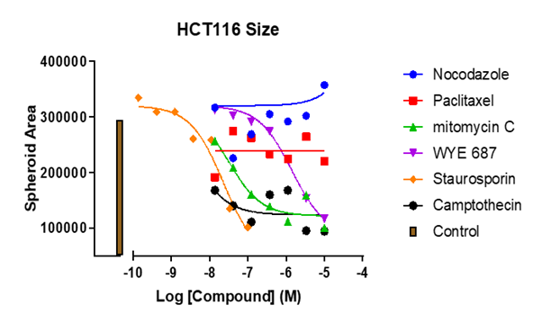

Figure 3: Spheroid area (in pixels) versus compound concentration in U87 (left) or HCT116 (middle) cells, taken from 8-day. Positive control: Cytotoxic effects of staurosporine can be observed between days 4 and 8 (right).
All compounds demonstrated activity in spheroids generated from U87 cells. While other compounds showed activity in spheroids generated from HCT116 cells, nocodazole and paclitaxel did not showed activity in spheroids from HCT116 spheroids.
Nocodazole
Paclitaxel
Mitomycin C
WYE 687
Sturoprine
Camptothecin
U87
154 nM
26 nM
177 nM
315 nM
21 nM
26 nM
HCT116
–
–
31 nM
1407 nM
23 nM
4 nM
Table 2: Estimated IC50 values derived from spheroid size at day 8.
Spheroid Viability


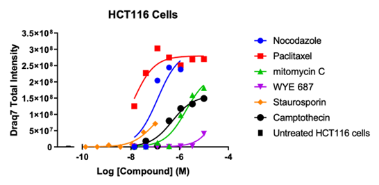
Figure 6: Graphical representation of the high content imaging data. DRAQ7TM staining of U87 spheroids (left) or HCT116 spheroids (right) at day 8 +/- compounds. Total intensity plotted versus dose response for each of the compounds tested. Note, some IC50 values were not plotted in Table 3 as they did not achieve 50% value at the maximum or minimum concentration used.
Nocodazole
Paclitaxel
Mitomycin C
WYE 687
Sturoprine
Camptothecin
U87
476 nM
233 nM
2107 nM
–
–
219 nM
HCT116
54 nM
125 nM
1857 nM
–
–
495 nM
Summary
All compounds tested affected cell viability in a dose-dependent manner (CellTiter Glo® 3D Viability Assay). Further investigation with a DRAQ7TM revealed dead cell stain for compounds that also showed reduced viability and a necrotic core. This effect was dose dependent.
Growth inhibition was determined by calculating spheroid area. We monitored growth inhibition in a dose- and time- dependent manner in both spheroid types. Taken together, we were able to build a pharmacological profile for each compound tested, and demonstrate that some test molecules showed increased efficacy under different pH conditions within the spheroid core.