As drug discovery programs evolve, the analysis of efficacy, pharmacokinetics, and pharmacodynamics becomes critical for lead optimization. Here we look at how Charnwood Discovery can streamline this process.
Outlining the preparation of animal study samples, xenograft models, and patient samples for analysis within our integrated departments. The key benefit of this setup is that it allows monitoring of signaling pathway activity and protein biomarker levels in Bioscience while evaluating compound concentrations in DMPK. The result is a powerful diagnostic tool to assess therapeutic effects.
Ex Vivo Sample Preparation
How do we go about sample preparation?
Beginning with the Precellys® Evolution Touch tissue homogenizer, which we use because of its efficiency and versatility for sample prep. This homogenizer uses glass or metal beads to grind, lyse, or homogenize a wide range of tissues, accommodating various tube sizes and even the most challenging samples. When coupled with the Cryolyos® Evolution, which cools samples down to 0˚C, it becomes ideal for biomarker analysis and the preparation of sensitive samples.
Within ADME / DMPK samples are homogenized in a buffer such as PBS and analyzed against a matrix-matched standard curve to determine compound concentration. A Thermo Scientific™ Exploris 120 Quadrupole Orbitrap mass spectrometer (MS) with a Thermo Scientific Vanquish Horizon ultra-high performance liquid chromatography (UHPLC) system is used for this.

In conjunction with biology the same tissue can be prepared in lysis buffer ready for biomarker analysis. Here we show how tissue samples analyzed with Simple Western™ Jess immunoblotting to evaluate target protein levels.
Samples are first homogenized, then a fixed quantity of protein, determined by BCA, is loaded and analyzed with specific antibodies. We have extensive experience preparing various tissue types, including liver, lungs, brain, muscle, kidney, and tumors, which are subsequently analyzed in immunoblotting assays.
This integrated approach ensures precise and comprehensive analysis of both protein targets and compound concentrations.
Analysis of Phospho-Proteins in Glucose-Challenged Rats
How does biomarker analysis plays an important role?
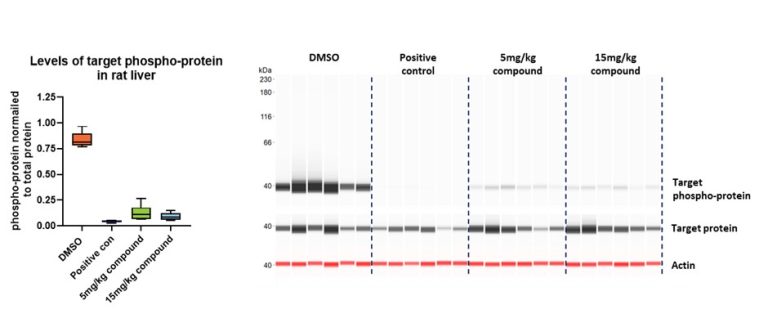
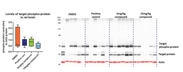
Determining whether a new drug reaches the relevant tissue at a therapeutic dose and achieves the desired biological effect is crucial for advancing a drug discovery project toward clinical trials. By harvesting the liver and brain from rats subjected to a glucose challenge, biomarker analysis was used to assess protein phosphorylation levels within the signaling cascade under investigation.
After establishing an optimized homogenization and lysis protocol, the samples were processed using Simple Western Jess and probed for target phospho-protein levels. The results demonstrated that the drug was highly effective in the liver (Figure 1) but did not alter biomarker levels in the brain (Figure 2). This discrepancy highlights the importance of tissue-specific analysis in evaluating drug efficacy and guiding further development.
Integrated Approach at Charnwood Discovery
At Charnwood Discovery, we take an integrated and meticulous approach to sample preparation and analysis. This ensures that we can effectively navigate the complexities of drug discovery, driving projects forward with precision and reliability.